Craniofacial defects such as cleft lip and cleft palate are among the most common birth defects. Lakshmi Kollara Sunil, assistant professor in the School of Communication Sciences and Disorders, is improving the way these patients are cared for by demonstrating the benefits of a team-based approach that pulls together all the experts needed to effectively treat these patients.
A cleft palate is a split or opening in the roof of the mouth, resulting in difficulties with feeding, speech, and resonance, or the quality of the voice that results from sound vibrations in the throat, mouth, and nose). Cleft lip, with or without cleft palate, affects one in 700 babies annually.
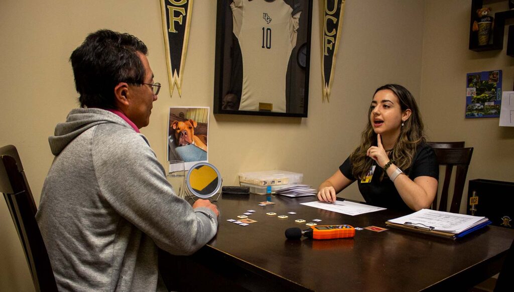
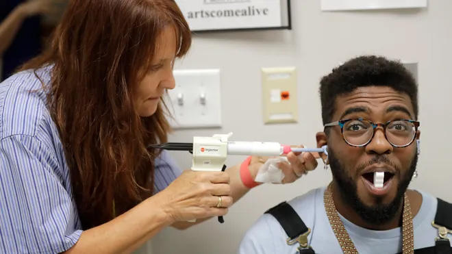
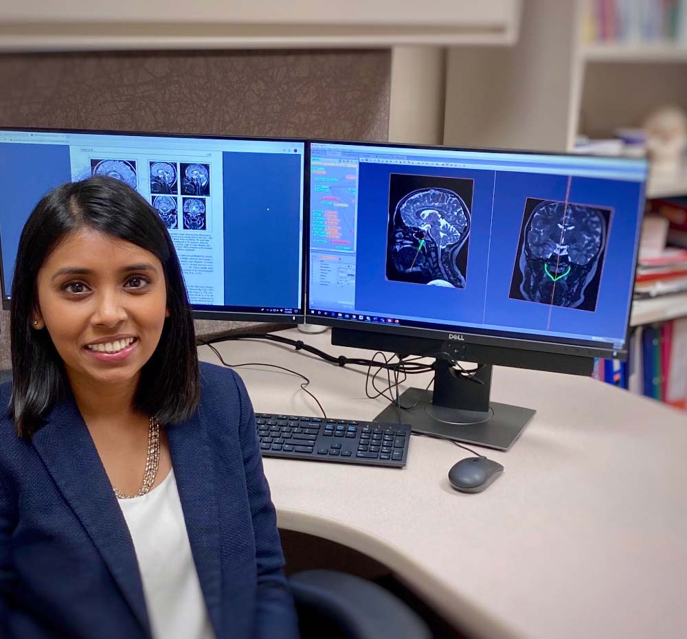
As director of the UCF Craniofacial and Speech Imaging Lab, Kollara’s research utilizes MRI and advanced imaging technology to improve the diagnosis, clinical management, and quality of life of individuals affected by cleft palate and other craniofacial anomalies.
Kollara first got interested in cleft palate after going on a cleft palate surgical mission trip to Mexico and through her research lab experiences as a graduate student.
“I enjoyed the multidisciplinary team approach in cleft palate care and enjoyed working with other healthcare professionals with the common goal of providing the best possible outcomes to our patients,” she said.
This collaborative team-based approach is something she is teaching to UCF students. She provides opportunities for students to learn from other cleft lip and cleft palate experts, such as surgeons, dentists, and psychologists . By learning with and from these experts, Kollara says speech-language pathologists become better clinicians in understanding and helping their patients.
Current collaborative research in the UCF Craniofacial and Speech Imaging Lab is focused on combining MRI and 3D imaging technology to study the speech mechanism in children with 22q11.2 deletion syndrome (22qDS), also commonly referred to as velocardiofacial syndrome or DiGeorge syndrome. The condition is the most common cause of syndromic palatal anomalies and velopharyngeal dysfunction. In children with 22qDS, there are variations of the velopharyngeal port that are still not well understood, which complicates surgery and results in less optimal post-surgical speech outcomes. By approaching this condition as a team, before the surgery is even done, Kollara hopes that they can improve patient outcomes.
“I believe that with this line of research, our field will see a major shift in the diagnostic and treatment procedures used for children with cleft palate and resonance disorders,” Kollara said.
Kollara says that she sees the advantages to the team-based approach in caring for this special population.
“Often, people think that a speech therapists’ work starts after the surgery,” said Kollara. “But our research is investigating new ways of providing comprehensive care right from the start in order to give patients the best outcomes possible.”